THE DNA
DNA, or deoxyribonucleic acid, is a molecule that carries the genetic instructions necessary for the growth, development, functioning, and reproduction of all known living organisms. DNA is a type of nucleic acid, and it plays a fundamental role in genetics and molecular biology. Here are some key points about DNA:
The polynucleotide chain is a fundamental component of DNA and RNA, and it is composed of a linear sequence of nucleotides. Each nucleotide in the chain consists of three main components: a phosphate group, a deoxyribose (in DNA) or ribose (in RNA) sugar molecule, and a nitrogenous base. The way these components are connected gives rise to the structure of the polynucleotide chain.
Phosphate Group: At one end of the nucleotide, there is a phosphate group (PO4). The phosphate group is a charged molecule that carries a negative charge. In the polynucleotide chain, the phosphate group of one nucleotide is covalently linked to the deoxyribose or ribose sugar of the next nucleotide, forming a sugar-phosphate backbone.
Sugar Molecule (Deoxyribose or Ribose): The sugar molecule in DNA is deoxyribose, while in RNA, it is ribose. Deoxyribose and ribose are five-carbon sugars. The carbon atoms in the sugar are numbered 1′ (1 prime), 2′, 3′, 4′, and 5′. The sugar is linked to the phosphate group at the 5′ carbon atom and to the nitrogenous base at the 1′ carbon atom. The 2′ carbon in the sugar differs between deoxyribose and ribose, with deoxyribose lacking an oxygen atom at the 2′ position.
Nitrogenous Base: The nitrogenous base is attached to the 1′ carbon of the sugar. There are four types of nitrogenous bases found in DNA and RNA:
- In DNA: Adenine (A), Thymine (T), Cytosine (C), and Guanine (G).
- In RNA: Adenine (A), Uracil (U), Cytosine (C), and Guanine (G).
The nitrogenous bases are responsible for the genetic code and base pairing. Adenine (A) pairs with thymine (T) in DNA or uracil (U) in RNA, and cytosine (C) pairs with guanine (G).
Sugar-Phosphate Backbone: The sugar-phosphate backbone is formed by the repeating linkage of phosphate groups and sugar molecules along the length of the polynucleotide chain. The backbone runs in the 5′ to 3′ direction, referring to the orientation of the carbon atoms in the sugar ring.
Base Pairing: In a double-stranded DNA molecule, the two polynucleotide chains are held together by hydrogen bonds between complementary nitrogenous bases. Adenine (A) pairs with thymine (T), and guanine (G) pairs with cytosine (C).
The salient features of the Double-helix structure of DNA are as follows:
Two Polynucleotide Chains: DNA consists of two polynucleotide chains, often referred to as strands. The sugar-phosphate backbone of each strand is on the outside, while the nitrogenous bases project inward.
Anti-Parallel Polarity: The two polynucleotide chains have anti-parallel polarity, meaning they run in opposite directions. If one chain has a 5′ to 3′ polarity, the other runs in the 3′ to 5′ direction.
Base Pairing and Hydrogen Bonds: The bases in the two strands are paired through hydrogen bonds. Adenine (A) always pairs with thymine (T) through two hydrogen bonds, and guanine (G) always pairs with cytosine (C) through three hydrogen bonds. This specific base pairing ensures complementary binding between the strands.
Right-Handed Helix: The double helix structure is coiled in a right-handed fashion. The helix has a pitch of approximately 3.4 nanometers (nm), and there are approximately 10 base pairs in each turn of the helix. This results in a distance of approximately 0.34 nm between each base pair.
Stacking of Base Pairs: The plane of one base pair stacks over the other in the double helix. This stacking, in addition to hydrogen bonds, contributes to the stability of the helical structure.
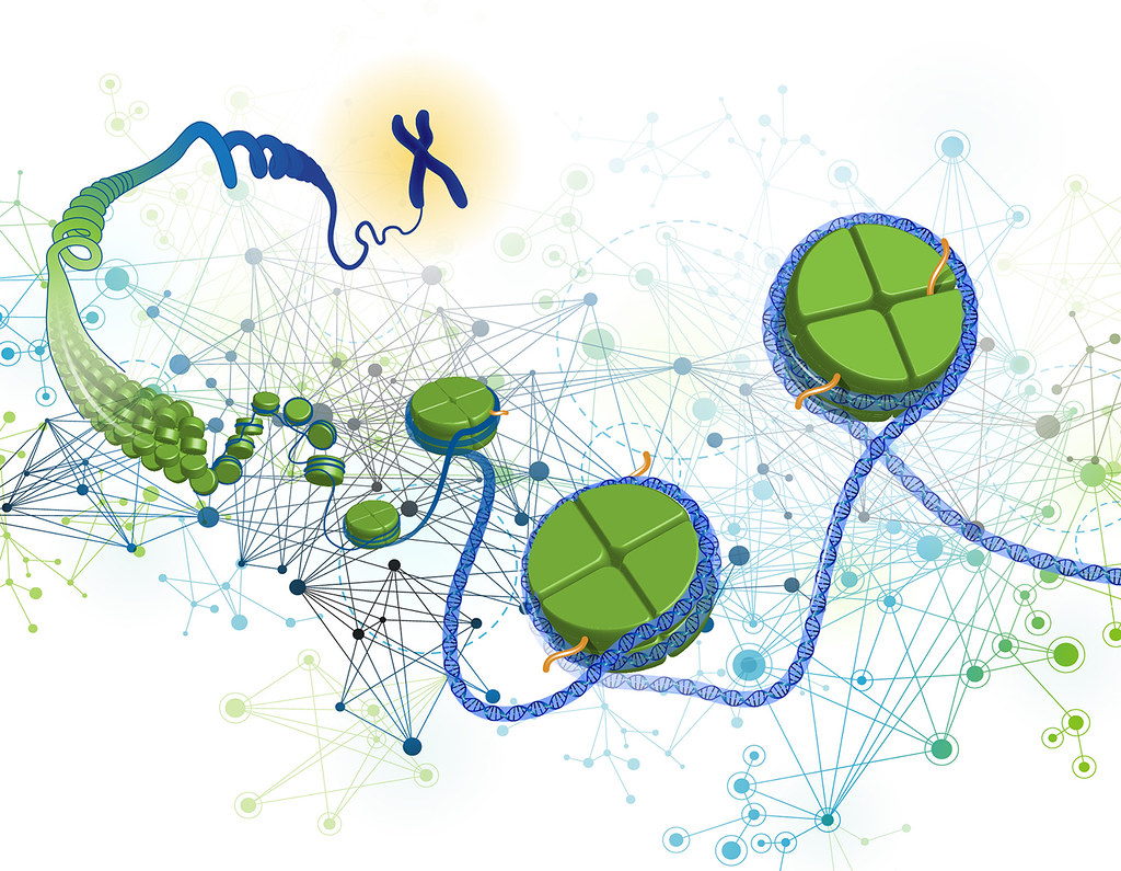
Packaging of DNA Helix
The packaging of DNA into a compact, organized structure is a critical aspect of genome function in cells. DNA in eukaryotic cells is much longer than the cell itself, so it must be efficiently packed to fit within the nucleus. Several levels of DNA packaging help achieve this compaction while still allowing for accessibility when needed. The primary levels of DNA packaging are:
Double Helix: The first level of DNA packaging is the formation of the double helix itself. DNA strands wrap around each other, forming the characteristic double-stranded structure.
Histones: DNA is associated with small, positively charged proteins called histones. DNA wraps around histones to form nucleosomes, the basic repeating unit of chromatin. Each nucleosome consists of about 147 base pairs of DNA coiled around an octamer of histone proteins. This helps condense DNA into a more organized structure.
Chromatin: The next level of DNA packaging involves the coiling of nucleosomes into a more complex structure known as chromatin. Chromatin consists of nucleosomes stacked on top of each other, and it can adopt different levels of compaction, depending on the cell’s needs. The less compacted form, called euchromatin, is associated with active gene expression. The highly compacted form, called heterochromatin, is associated with gene silencing and overall compaction.
- Beads-on-String Structure: The basic unit of chromatin is the nucleosome, where DNA wraps around a core of histone proteins, forming a “beads-on-string” structure. This structure allows for the compaction of DNA and also regulates access to genetic information.
Chromosomes: The highest level of DNA packaging involves the condensation of chromatin into distinct chromosomes. During cell division, chromatin further compacts to form highly visible structures known as chromosomes. Each chromosome contains a single, long DNA molecule that has been tightly coiled and condensed to allow for accurate segregation during cell division.
Metaphase Chromosomes: During cell division (metaphase), chromatin fibers condense further to form highly visible and condensed structures known as chromosomes. This is necessary for the accurate segregation of genetic material into daughter cells.
Non-Histone Chromosomal (NHC) Proteins: While histones are crucial for the organization of DNA into nucleosomes, NHC proteins play a role in packaging chromatin at higher levels. These proteins help in coiling and condensing chromatin fibers into more compact structures, including the formation of metaphase chromosomes.
Euchromatin and Heterochromatin: The chromatin in the nucleus can exist in two main states—euchromatin and heterochromatin. Euchromatin is loosely packed, stains light, and is associated with active gene expression. In contrast, heterochromatin is more densely packed and stains dark, and it is generally transcriptionally inactive. The distinction between these two states reflects the accessibility of the genetic information within the chromatin.

