WHAT IS A CELL?
- Definition: The cell is the fundamental structural and functional unit of all living organisms.
- Attributes of Unicellular Organisms: Capable of independent existence and performing essential life functions.
- Historical Perspective:
- Anton Von Leeuwenhoek: First to observe and describe live cells.
- Robert Brown: Discovered the cell nucleus.
- Microscopy Advancements: Invention and improvement, including electron microscopy, revealed detailed cell structures.
CELL THEORY
- Origins: Developed by Matthias Schleiden and Theodore Schwann in the 19th century.
- Context: Examination of plant and animal cells led to the formulation of fundamental principles.
I. Contributors to Cell Theory:
- Matthias Schleiden (1838):
- German botanist.
- Studied plants and identified different cell types composing plant tissues.
- Theodore Schwann (1839):
- British Zoologist.
- Investigated animal cells, identified the plasma membrane, and proposed the presence of cell walls in plant cells.
- Hypothesized that the bodies of animals and plants are composed of cells and their products.
- Rudolf Virchow (1855):
- German physician.
- Proposed “Omnis cellula-e cellula” (Every cell arises from a pre-existing cell).
- Modified and finalized the cell theory by emphasizing cell division and the origin of new cells.
II. Cell Theory Principles:
- First Principle:
- All living organisms are composed of cells and their products.
- Second Principle:
- All cells arise from pre-existing cells.
- Rudolf Virchow’s Contribution: Clarified the mechanism of cell division and the continuity of cellular life.
AN OVERVIEW OF CELL
- Observation of Cells:
- Microscopic examination of onion peel and human cheek cells.
- Recollection of cell structures.
I. Basic Cell Structure:
- Plant Cell (Onion):
- Cell Wall: Outer boundary, providing structural support.
- Cell Membrane: Lies just within the cell wall.
- Nucleus: Dense, membrane-bound structure containing chromosomes with DNA.
- Animal Cell (Cheek):
- Cell Membrane: Outer delimiting structure.
- Nucleus: Membrane-bound structure containing genetic material.
II. Cell Types:
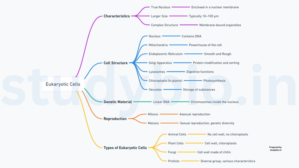
- Eukaryotic Cells:
- Definition: Cells with membrane-bound nuclei.
- Examples: Plant and animal cells.
- Prokaryotic Cells:
- Definition: Cells lacking a membrane-bound nucleus.
- Examples: Bacteria.
III. Cellular Components:
- Cytoplasm:
- Description: Semi-fluid matrix occupying cell volume.
- Function: A main arena for cellular activities; site of various chemical reactions.
- Organelles (Eukaryotic Cells):
- Endoplasmic Reticulum (ER), Golgi Complex, Lysosomes, Mitochondria, Microbodies, Vacuoles.
- Ribosomes:
- Distribution: Found in cytoplasm, chloroplasts, mitochondria, and on rough ER.
- Centrosome (Animal Cells):
- Function: Aids in cell division.
IV. Cell Size, Shape, and Diversity:
- Size Variation:
- Examples: Mycoplasmas (0.3 µm), bacteria (3-5 µm), human red blood cells (7.0 µm), ostrich egg (largest isolated single cell).
- Shape Variation:
- Examples: Disc-like, polygonal, columnar, cuboid, thread-like, irregular.
- Functional Dependence: Cell shape may vary based on the function it performs.
PROKARYOTIC CELLS
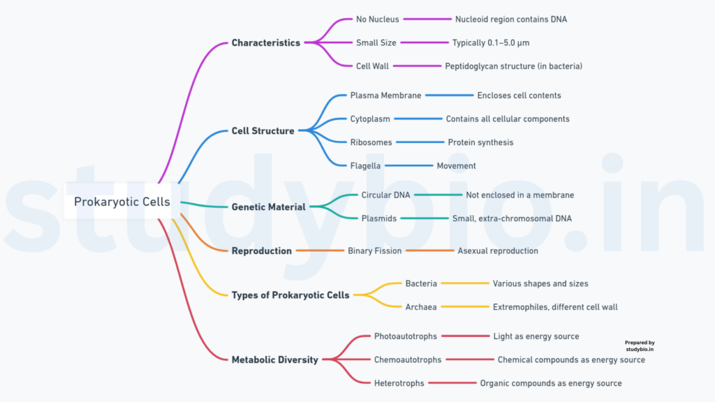
Introduction:
- Representation:
- Bacteria, blue-green algae, mycoplasma, and PPLO (Pleuro Pneumonia Like Organisms).
- Characteristics:
- Generally smaller and faster multiplication compared to eukaryotic cells.
- Varying shapes and sizes.
I. Cell Shapes:
- Basic Shapes of Bacteria:
- Bacillus: Rod-like.
- Coccus: Spherical.
- Vibrio: Comma-shaped.
- Spirillum: Spiral.
II. Organization of Prokaryotic Cells:
- Cell Wall:
- Surrounds the cell membrane, present in all prokaryotes except mycoplasma.
- Cytoplasm:
- Fluid matrix filling the cell.
- Nucleus:
- Not well-defined; genetic material is naked.
- Genomic DNA:
- Single chromosome/circular DNA.
- Plasmids:
- Small circular DNA outside the genomic DNA; confers unique phenotypic characters, e.g., antibiotic resistance.
- Organelles:
- Absence of membrane-bound organelles except for ribosomes.
- Inclusions:
- A unique feature in the form of a specialized differentiated cell membrane called mesosome; is the infoldings of the cell membrane.
Cell Envelope and its Modifications
- Cell Envelope:
- Composition:
- Outermost glycocalyx, cell wall, and plasma membrane.
- Collective Function:
- Acts as a single protective unit.
- Composition:
- Classification:
- Gram-positive: Takes up the gram stain.
- Gram-negative: Does not take up the gram stain.
I. Components of Cell Envelope:
- 1. Glycocalyx:
- Types:
- Slime layer (loose sheath).
- Capsule (thick and tough).
- Function:
- Varies in composition and thickness; provides protection.
- Types:
- 2. Cell Wall:
- Role:
- Determines cell shape and provides structural support.
- Role:
- 3. Plasma Membrane:
- Characteristics:
- Selectively permeable.
- Structurally similar to eukaryotic membranes.
- Characteristics:
II. Special Membranous Structures:
- 1. Mesosome:
- Formation:
- Extensions of plasma membrane into the cell.
- Functions:
- Aids in cell wall formation, DNA processes, respiration, and secretion.
- Formation:
- 2. Chromatophores:
- Presence:
- Found in cyanobacteria.
- Membranous extensions containing pigments.
- Presence:
III. Motility in Bacterial Cells:
- Flagella:
- Structure:
- Composed of filament, hook, and basal body.
- Variation:
- Range in the number and arrangement.
- Structure:
- Surface Structures:
- 1. Pili:
- Elongated tubular structures are made of a special protein.
- 2. Fimbriae:
- Small bristle-like fibers sprouting from the cell.
- Aid in attachment to surfaces and host tissues.
- 1. Pili:
Ribosomes and Inclusion Bodies
I. Ribosomes in Prokaryotes:
- Association:
- Associated with the plasma membrane.
- Size and Structure:
- About 15 nm by 20 nm.
- Composed of 50S and 30S subunits.
- Together form 70S prokaryotic ribosomes.
- Function:
- Site of protein synthesis.
- Polyribosomes:
- A chain is formed when several ribosomes attach to a single mRNA.
- Facilitates simultaneous protein synthesis.
II. Inclusion Bodies:
- Nature:
- Reserve material stored in the cytoplasm.
- Membrane Association:
- Not bound by any membrane system.
- Lie freely in the cytoplasm.
- Examples:
- Phosphate granules.
- Cyanophycean granules.
- Glycogen granules.
- Gas Vacuoles:
- Found in blue-green and purple-green photosynthetic bacteria.
EUKARYOTIC CELLS
The cell membrane, a crucial component of cells, was extensively studied with the advent of the electron microscope in the 1950s. Chemical studies, particularly in human red blood cells (RBCs), provided insights into its composition. The cell membrane primarily consists of lipids and proteins, with phospholipids forming a bilayer. The arrangement places the polar heads outward and the hydrophobic tails inward, protecting them from the aqueous environment. Cholesterol is also present in the membrane.
1. Composition:
- Lipids:
- Majorly phospholipids.
- Arranged in a bilayer.
- Cholesterol is present.
- Proteins:
- Integral and peripheral proteins.
- Integral proteins partially or totally embedded.
- Carbohydrates:
- Present in the membrane.
2. Fluid Mosaic Model:
- Proposed by Singer and Nicolson in 1972.
- Describes the membrane as a quasi-fluid structure.
- Enables lateral movement of proteins within the bilayer.
- Fluidity is crucial for cell functions like growth, intercellular junction formation, secretion, and cell division.
3. Functions:
- Selective Permeability:
- Allows certain molecules to pass through.
- Passive Transport:
- Movement without energy requirement.
- Includes simple diffusion and osmosis.
- Active Transport:
- Energy-dependent process.
- Involves ATP utilization.
- Example: Na+/K+ Pump.
Cell Wal
The cell wall, a non-living structure, envelops the plasma membrane in fungi and plants. It serves various crucial functions, including providing structural support, protection against mechanical damage and infections, facilitating cell-to-cell interactions, and acting as a barrier to unwanted macromolecules.
1. Composition:
- Plants:
- Primary components include cellulose, hemicellulose, pectins, and proteins.
- The secondary wall forms as the cell matures.
- Algae:
- The cell wall comprises cellulose, galactans, mannans, and minerals like calcium carbonate.
- Fungi:
- Contains chitin, glucans, and proteins.
2. Functions:
- Structural Support:
- Gives shape to the cell.
- Maintains cell integrity.
- Protection:
- Shields against mechanical damage and infections.
- Cell-to-Cell Interaction:
- Facilitates communication between adjacent cells.
- Barrier:
- Prevents entry of undesirable macromolecules.
3. Growth and Maturation:
- Primary Wall:
- Capable of growth.
- Diminishes as the cell matures.
- Secondary Wall:
- Forms on the inner side of the cell membrane in mature cells.
4. Middle Lamella:
- Composition:
- Mainly calcium pectate.
- Function:
- Holds neighboring cells together.
- Forms a gluing layer.
5. Plasmodesmata:
- Structure:
- Channels.
- Function:
- Traverse cell wall and middle lamella.
- Connect cytoplasm of neighboring cells.
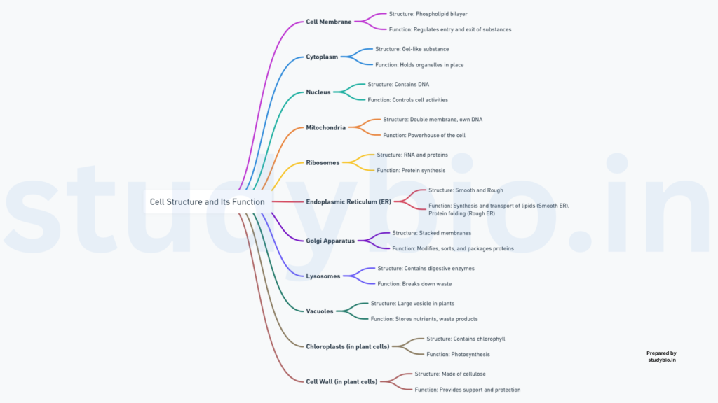
Endomembrane System
ENDOPLASMIC RETICULUM (ER)
The endoplasmic reticulum (ER) is a network of tiny tubular structures found in the cytoplasm of eukaryotic cells. It plays a crucial role in various cellular processes and is divided into two compartments: luminal (inside ER) and extraluminal (cytoplasm) compartments.
1. Types of Endoplasmic Reticulum:
- Rough Endoplasmic Reticulum (RER):
- Appearance:
- Ribosomes attached to the outer surface.
- Function:
- Actively involved in protein synthesis and secretion.
- Extensive and continuous with the outer membrane of the nucleus.
- Appearance:
- Smooth Endoplasmic Reticulum (SER):
- Appearance:
- Lacks ribosomes, appearing smooth.
- Function:
- A major site for lipid synthesis.
- Synthesizes lipid-like steroidal hormones in animal cells.
- Appearance:
2. Functions of Endoplasmic Reticulum:
- Protein Synthesis:
- RER is involved in the synthesis and secretion of proteins.
- Lipid Synthesis:
- SER is the primary site for lipid synthesis.
- Steroid Hormone Synthesis:
- SER in animal cells synthesizes steroidal hormones.
- Transport and Storage:
- Facilitates the transport of synthesized molecules.
- Stores synthesized molecules.
3. Structure and Appearance:
- Tubular Network:
- Microscopic tubular structures are scattered in the cytoplasm.
- Rough Appearance:
- Ribosomes attached to the outer surface.
- Smooth Appearance:
- Lacks ribosomes, appearing smooth.
4. Role in Cellular Processes:
- Protein Secretion:
- RER facilitates the synthesis and secretion of proteins.
- Lipid Metabolism:
- SER is involved in lipid metabolism and synthesis.
- Detoxification:
- SER plays a role in detoxifying drugs and poisons.
GOLGI APPARATUS
The Golgi apparatus, named after Camillo Golgi who first observed its structures, is a cellular organelle consisting of flat, disc-shaped sacs or cisternae with a diameter ranging from 0.5µm to 1.0µm. These cisternae are stacked parallel to each other and are organized near the nucleus. The Golgi apparatus has distinct convex (cis) and concave (trans) faces, with varied numbers of cisternae forming a Golgi complex.
1. Structure of Golgi Apparatus:
- Cisternae:
- Flat, disc-shaped sacs stacked parallel to each other.
- Diameter: 0.5µm to 1.0µm.
- Faces:
- Convex cis face (forming face).
- Concave trans face (maturing face).
- Arrangement:
- Concentric arrangement near the nucleus.
2. Function of Golgi Apparatus:
- Packaging and Modification:
- Packages materials for delivery to intra-cellular targets or secretion outside the cell.
- Vesicles from the endoplasmic reticulum (ER) fuse with the cis face and move towards the maturing face.
- Works closely with the endoplasmic reticulum (ER).
- Protein Modification:
- Modifies proteins synthesized by ribosomes on the ER.
- Important for the modification and processing of proteins before release from the trans face.
- Glycoprotein and Glycolipid Formation:
- Key site for the formation of glycoproteins and glycolipids.
3. Interconnected Faces:
- Cis and Trans Faces:
- Entirely different but interconnected.
- Cis face receives materials, and trans face releases modified and packaged products.
4. Association with Endoplasmic Reticulum (ER):
- Close Association:
- Golgi apparatus and ER work in close association.
- Materials move from the ER to the Golgi apparatus for modification and packaging.
LYSOSOMES
Lysosomes are membrane-bound vesicular structures that originate through the packaging process in the Golgi apparatus. These vesicles are characterized by their high concentration of various hydrolytic enzymes, collectively known as hydrolases. Lysosomes play a crucial role in intracellular digestion and the recycling of cellular components.
1. Formation and Origin:
- Golgi Apparatus:
- Formed through the packaging process in the Golgi apparatus.
- Membrane-bound vesicles.
2. Enzyme Content:
- Hydrolytic Enzymes:
- Rich in a variety of hydrolytic enzymes (hydrolases).
- Lipases (digest lipids).
- Proteases (digest proteins).
- Carbohydrases (digest carbohydrates).
- Enzymes are optimally active at acidic pH.
- Rich in a variety of hydrolytic enzymes (hydrolases).
3. Functions of Lysosomes:
- Intracellular Digestion:
- Responsible for breaking down various biomolecules within the cell.
- Digestion of carbohydrates, proteins, lipids, and nucleic acids.
- Autophagy:
- Involves the digestion of worn-out organelles and cellular debris.
- Recycling of cellular components.
4. Acidic Environment:
- pH Optimum:
- Enzymes within lysosomes are optimally active at acidic pH.
- Acidic environment aids in the efficient hydrolysis of macromolecules.
5. Versatility in Digestive Functions:
- Carbohydrates, Proteins, Lipids, Nucleic Acids:
- Capable of digesting a wide range of biomolecules:
- Carbohydrates.
- Proteins.
- Lipids.
- Nucleic acids.
- Capable of digesting a wide range of biomolecules:
VACUOLES
Vacuoles are membrane-bound organelles found in the cytoplasm, enclosed by a single membrane called the tonoplast. These structures are present in various types of cells, with notable prominence in plant cells. Vacuoles serve multiple functions, including storage of water, sap, and waste products, contributing to the overall structure and function of the cell.
1. Structure and Composition:
- Membrane-Bound Space:
- Vacuoles are membrane-bound spaces within the cytoplasm.
- Tonoplast:
- Enclosed by a single membrane known as tonoplast.
2. Prominence in Plant Cells:
- Volume Occupancy:
- In plant cells, vacuoles can occupy a significant portion, up to 90%, of the cell volume.
- Function in Plants:
- Essential for maintaining turgor pressure.
- Storage of water, ions, and other substances.
3. Transport Functions:
- Ion and Material Transport:
- Tonoplast facilitates the transport of ions and various materials into the vacuole.
- Concentrations of substances in the vacuole can be significantly higher than in the cytoplasm.
4. Roles in Different Organisms:
- Amoeba:
- Contractile Vacuole:
- Important for osmoregulation and excretion.
- Contractile Vacuole:
- Protists:
- Food Vacuoles:
- Formation by engulfing food particles.
- Food Vacuoles:
5. Functions:
- Storage:
- Storage of water, sap, and other substances not immediately useful to the cell.
- Maintaining Turgor Pressure:
- Crucial for plant cells to maintain structural integrity and rigidity.
- Waste Storage:
- Accumulation of excretory products and materials.
MITOCHONDRIA
Mitochondria are essential double membrane-bound organelles involved in aerobic respiration, commonly known as the “powerhouses” of the cell. These structures generate cellular energy in the form of ATP and exhibit variability in terms of shape, size, and number depending on the physiological activity of the cell.
1. Structural Characteristics:
- Shape and Size:
- Typically sausage-shaped or cylindrical.
- Diameter: 0.2-1.0µm (average 0.5µm).
- Length: 1.0-4.1µm.
- Double Membrane Structure:
- Outer membrane: Forms the continuous boundary.
- Inner membrane: Contains infoldings known as cristae, increasing surface area.
- Compartments:
- Outer Compartment.
- Inner Compartment: Filled with a dense homogeneous substance called the matrix.
2. Function:
- Aerobic Respiration:
- Mitochondria are the primary sites for aerobic respiration.
- Production of ATP (Adenosine Triphosphate).
3. Components:
- Matrix:
- Dense homogeneous substance filling the inner compartment.
- Genetic Material:
- Single circular DNA molecule.
- Few RNA molecules.
- Ribosomes (70S).
- Enzymes:
- Specific enzymes associated with mitochondrial function.
4. Physiological Variability:
- Number:
- Variable per cell based on physiological activity.
- Shape and Size Variability:
- Considerable degree of variability observed.
5. Reproduction:
- Fission:
- Mitochondria reproduce through fission.
PLASTIDS
Plastids are organelles found in plant cells and euglenoids, contributing to the characteristic colors of plants due to specific pigments. Plastids can be categorized into chloroplasts, chromoplasts, and leucoplasts based on their pigment content.
1. Types of Plastids:
- Chloroplasts:
- Contain chlorophyll and carotenoid pigments.
- Responsible for trapping light energy during photosynthesis.
- Chromoplasts:
- Contain fat-soluble carotenoid pigments (e.g., carotene, xanthophylls).
- Impart yellow, orange, or red color to plant parts.
- Leucoplasts:
- Colorless plastids with varied shapes and sizes.
- Store nutrients:
- Amyloplasts: Store carbohydrates (starch), e.g., in potatoes.
- Elaioplasts: Store oils and fats.
- Aleuroplasts: Store proteins.
2. Distribution and Characteristics:
- Localization:
- Found in all plant cells, especially abundant in mesophyll cells of leaves.
- Variability:
- Shapes: Lens-shaped, oval, spherical, discoid, or ribbon-like.
- Size: 5-10µm in length, 2-4µm in width.
- Number: Varies from 1 (e.g., in Chlamydomonas) to 20-40 per cell in mesophyll.
- Membrane Structure:
- Double membrane-bound organelles.
- The inner membrane is less permeable.
- Internal Structures:
- Stroma:
- Space is limited by the inner membrane, containing enzymes for synthesis.
- Thylakoids:
- Organized, flattened membranous sacs in the stroma.
- Form stacks called grana (singular: granum) or intergranal thylakoids.
- Stroma Lamellae:
- Flat membranous tubules connecting thylakoids of different grana.
- Lumen:
- Space is enclosed by the thylakoid membrane.
- Stroma:
- Content:
- Enzymes for carbohydrate and protein synthesis.
- Small, double-stranded circular DNA molecules and ribosomes.
- Chlorophyll pigments in thylakoids.
- Smaller ribosomes (70S) compared to cytoplasmic ribosomes (80S).
RIBOSOMES
Ribosomes are essential cellular structures responsible for protein synthesis. Discovered by George Palade in 1953 under the electron microscope, ribosomes consist of ribonucleic acid (RNA) and proteins and lack a surrounding membrane.
1. Types of Ribosomes:
- Eukaryotic Ribosomes (80S):
- Composed of two subunits: 60S (larger) and 40S (smaller).
- Prokaryotic Ribosomes (70S):
- Composed of two subunits: 50S and 30S.
2. Composition and Structure:
- Subunit Composition:
- Both 70S and 80S ribosomes have two subunits each.
- The designation (e.g., 60S, 40S) represents the sedimentation coefficient (Svedberg’s Unit), indicating density and size indirectly.
- Size Difference:
- Eukaryotic ribosomes (80S) are larger than prokaryotic ribosomes (70S).
- Subunit Components:
- 80S Ribosomes:
- Larger Subunit (60S).
- Smaller Subunit (40S).
- 70S Ribosomes:
- Larger Subunit (50S).
- Smaller Subunit (30S).
- 80S Ribosomes:
3. Functional Significance:
- Ribosomes play a crucial role in protein synthesis, translating genetic information from mRNA into proteins.
- The assembly of amino acids into polypeptide chains occurs in the ribosomes.
CYTOSKELETON
The cytoskeleton is a dynamic network of filamentous protein structures within the cytoplasm, providing structural support and contributing to various cellular functions. It encompasses microtubules, microfilaments, and intermediate filaments.
1. Components of the Cytoskeleton:
- Microtubules:
- Hollow tubes made of tubulin protein subunits.
- Function in cell shape maintenance, intracellular transport, and cell division.
- Microfilaments (Actin Filaments):
- Thin filaments composed of actin protein subunits.
- Contribute to cell shape, muscle contraction, and cell motility.
- Intermediate Filaments:
- Intermediate in size compared to microtubules and microfilaments.
- Provide mechanical support and anchor organelles.
2. Functions of the Cytoskeleton:
- Mechanical Support:
- Maintains cell shape and rigidity.
- Supports structural integrity against external forces.
- Cell Motility:
- Facilitates cellular movement, including intracellular transport and migration.
- Microtubules serve as tracks for motor proteins.
- Intracellular Transport:
- Microtubules act as highways for vesicle and organelle movement.
- Motor proteins, such as dynein and kinesin, move along these structures.
- Cell Division:
- Guides the formation of the mitotic spindle during cell division.
- Ensures proper chromosome segregation.
3. Dynamic Nature:
- The cytoskeleton is dynamic, allowing cells to adapt to changing conditions.
- Reorganization of cytoskeletal elements is crucial for cell functions.
CILIA AND FLAGELLA
Cilia and flagella are specialized hair-like structures protruding from the cell membrane, serving essential roles in cell movement and fluid propulsion.
1. Structural Characteristics:
- Cilia:
- Small, numerous hair-like projections.
- Function like oars, causing movement of the cell or surrounding fluid.
- Flagella:
- Longer, responsible for cell movement.
- Commonly found as a single or a few projections.
2. Structural Composition:
- Both cilia and flagella are covered with a plasma membrane.
- Core structure, known as the axoneme, contains microtubules.
- Axoneme typically exhibits a 9+2 array:
- Nine doublets of radially arranged peripheral microtubules.
- A pair of centrally located microtubules.
- Bridges and linkers connect the microtubules.
3. Central Role of Basal Bodies:
- Cilia and flagella emerge from centriole-like structures called basal bodies.
- Basal bodies serve as the anchoring point for the assembly of axonemal microtubules.
4. Function and Movement:
- Cilia:
- Propel fluids over the cell surface.
- Found in large numbers on certain epithelial cells.
- Flagella:
- Propel the entire cell.
- Examples include the tail of sperm cells.
5. Prokaryotic vs. Eukaryotic Flagella:
- Prokaryotic bacteria possess flagella, structurally distinct from eukaryotic flagella.
6. Electron Microscopic Features:
- Cilia and flagella are visualized under electron microscopy.
- 9+2 array, radial spokes, and central sheath are distinctive features.
CENTROSOME AND CENTRIOLES
The centrosome, housing two cylindrical centrioles, is a crucial organelle involved in cellular organization and division. Here’s an in-depth exploration:
1. Centrosome Composition:
- Typically contains two cylindrical structures, the centrioles.
- Surrounded by a matrix of amorphous pericentriolar materials.
2. Centriole Structure:
- Orientation:
- Both centrioles within a centrosome lie perpendicular to each other.
- Cartwheel Organization:
- Structurally organized like a cartwheel.
- Composed of nine evenly spaced peripheral fibrils made of tubulin protein.
- Adjacent triplets are interconnected, forming a cohesive structure.
- Hub:
- Central part of the proximal region of the centriole.
- A proteinaceous hub connected to tubules of peripheral triplets by radial spokes.
3. Functional Roles:
- Basal Body Formation:
- Centrioles serve as the basal body for the formation of cilia or flagella.
- Spindle Fibres and Apparatus:
- Critical in the formation of spindle fibers during cell division in animal cells.
- Integral for the assembly of the spindle apparatus, facilitating chromosome segregation.
4. Microtubule Organization:
- Centrioles are crucial in the organization of microtubules within the cell.
- Microtubules radiate from the centrosome, contributing to cellular structure and movement.
5. Cell Division:
- Centrosomes play a pivotal role in orchestrating cell division.
- They contribute to the organization of the mitotic spindle, ensuring accurate segregation of chromosomes.
6. Importance in Cellular Organization:
- Centrosomes are central in maintaining cell structure and organization.
- They influence cytoskeletal dynamics, impacting cellular motility and shape.
NUCLEUS
The nucleus, a pivotal cell organelle, plays a central role in cellular functions and genetic regulation. Here’s a comprehensive overview:
1. Discovery and Components:
- First described by Robert Brown in 1831.
- Chromatin, is the material stained by basic dyes, named by Flemming.
- The interphase nucleus comprises:
- Elaborate nucleoprotein fibers called chromatin.
- Nuclear matrix containing nucleoli.
- One or more spherical bodies are known as nucleoli.
2. Nuclear Envelope and Pores:
- Consists of two parallel membranes with a perinuclear space (10 to 50 nm).
- Acts as a barrier between the nucleus and cytoplasm.
- Contains pores for the movement of RNA and protein molecules.
- Continuous with the endoplasmic reticulum, bearing ribosomes.
3. Variability in Nuclei:
- Normally one nucleus per cell, exceptions exist.
- Some organisms have more than one nucleus per cell.
- Certain mature cells lack a nucleus (e.g., erythrocytes, sieve tube cells).
- Consideration of “living” status in cells lacking a nucleus raises interesting questions.
4. Nuclear Matrix and Nucleoplasm:
- The nuclear matrix or nucleoplasm contains nucleoli and chromatin.
- Nucleoli are non-membrane-bound structures involved in active ribosomal RNA synthesis.
- Chromatin, a loose network of nucleoprotein fibers, becomes structured chromosomes during cell division.
5. Chromatin Composition:
- Comprises DNA, histones (basic proteins), non-histone proteins, and RNA.
- A single human cell contains approximately two meters of DNA distributed among its forty-six chromosomes.
6. Chromosome Structure:
- Chromosomes are visible only during cell division.
- Primary constriction or centromere holds two chromatids.
- Kinetochores are disc-shaped structures on the sides of the centromere.
- Classification based on centromere position:
- Metacentric: Middle centromere, forming two equal arms.
- Sub-metacentric: Centromere slightly away from the middle.
- Acrocentric: Centromere close to one end, forming short and long arms.
- Telocentric: Terminal centromere.
MICROBODIES
Microbodies, crucial membrane-bound organelles, contribute to diverse cellular functions through the containment of various enzymes. Here’s a detailed exploration:
1. Nature and Composition:
- Microbodies are membrane-bound vesicles found in both plant and animal cells.
- These vesicles encapsulate a variety of enzymes.
2. Functions:
- Diverse Enzymatic Activities: Microbodies house enzymes that participate in various metabolic activities crucial for cellular functions.
- Metabolism Regulation: Enzymes within microbodies contribute to the regulation of specific metabolic pathways.
3. Types of Microbodies:
- Peroxisomes: A type of microbody involved in processes like lipid metabolism and detoxification reactions. They contain enzymes like catalase.
- Glyoxysomes: Found in plant cells, particularly in germinating seeds. They facilitate the conversion of fats to sugars.
- Glycosomes: Present in some protists and are involved in glycolysis.
4. Peroxisomes:
- Lipid Metabolism: Peroxisomes play a crucial role in lipid metabolism, including the breakdown of fatty acids.
- Detoxification: Involved in detoxifying harmful substances, particularly hydrogen peroxide, through the action of catalase.
5. Glyoxysomes:
- Seed Germination: Glyoxysomes are prominent during the germination of seeds, aiding in the conversion of stored lipids into carbohydrates.
6. Significance in Cellular Health:
- Homeostasis: Microbodies contribute to cellular homeostasis by regulating specific metabolic pathways.
- Cellular Adaptation: The presence of microbodies reflects the adaptability of cells to different metabolic demands.
Pingback: Grade 11 Notes (Biology)