ENDOCRINE GLANDS AND HORMONES
Endocrine glands, devoid of ducts, are referred to as ductless glands, releasing hormones into the bloodstream. Hormones, acting as intercellular messengers, are produced in trace amounts. In the human endocrine system, various glands and tissues contribute to hormonal regulation.
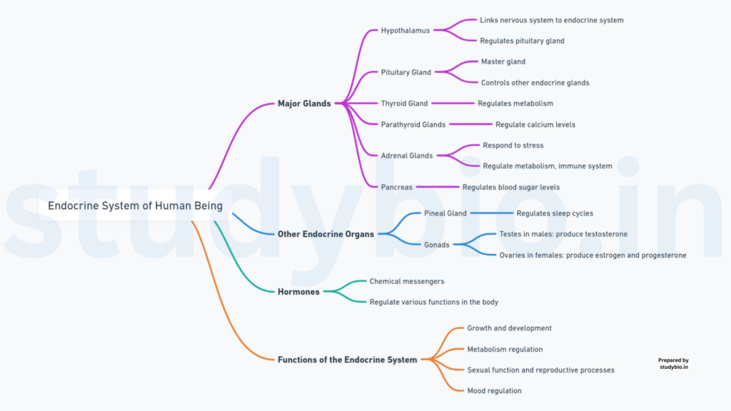
HUMAN ENDOCRINE SYSTEM
The endocrine system comprises organized endocrine glands and diffused hormone-producing tissues/cells dispersed throughout the body. Key components include:
- Pituitary Gland:
- Location: Situated at the base of the brain.
- Function: Regulates other endocrine glands; secretes growth hormone, thyroid-stimulating hormone, and more.
- Pineal Gland:
- Location: Located in the brain.
- Function: Produces melatonin, influencing sleep-wake cycles.
- Thyroid Gland:
- Location: Found in the neck.
- Function: Secretes thyroxine, controlling metabolism.
- Adrenal Glands:
- Location: Positioned atop each kidney.
- Function: Releases hormones like adrenaline, influencing the “fight or flight” response.
- Pancreas:
- Location: Lies behind the stomach.
- Function: Produces insulin and glucagon, regulating blood sugar levels.
- Parathyroid Glands:
- Location: Attached to the thyroid gland.
- Function: Maintains calcium balance in the body.
- Thymus:
- Location: Situated in the upper chest.
- Function: Vital for immune system development.
- Gonads (Testis/Ovary):
- Location: Testes in males; ovaries in females.
- Function: Regulates reproductive functions; produces sex hormones.
THE HYPOTHALAMUS
The hypothalamus, situated at the basal part of the diencephalon within the forebrain, plays a pivotal role in regulating diverse body functions (Figure 19.1). Comprising various neurosecretory cell groups called nuclei, the hypothalamus produces hormones that exert control over pituitary gland activities.
HYPOTHALAMIC HORMONES
The hormones generated by the hypothalamus fall into two categories:
- Releasing Hormones:
- Function: Stimulate the secretion of pituitary hormones.
- Example: Gonadotrophin-releasing hormone (GnRH) prompts the synthesis and release of gonadotrophins from the pituitary.
- Inhibiting Hormones:
- Function: Inhibit the secretion of pituitary hormones.
- Example: Somatostatin from the hypothalamus inhibits the release of growth hormone by the pituitary.
THE PITUITARY GLAND
The pituitary gland, ensconced in the bony cavity known as sella turcica, is tethered to the hypothalamus by a stalk. It anatomically comprises the adenohypophysis and the neurohypophysis.

Adenohypophysis:
- Pars Distalis (Anterior Pituitary):
- Hormones Produced:
- Growth Hormone (GH)
- Prolactin (PRL)
- Thyroid Stimulating Hormone (TSH)
- Adrenocorticotrophic Hormone (ACTH)
- Luteinizing Hormone (LH)
- Follicle Stimulating Hormone (FSH)
- Hormones Produced:
- Pars Intermedia:
- Hormone Produced:
- Melanocyte Stimulating Hormone (MSH)
- Hormone Produced:
Note: In humans, pars intermedia is often integrated with pars distalis.
Neurohypophysis (Posterior Pituitary):
- Hormones Stored and Released:
- Oxytocin
- Vasopressin (Antidiuretic Hormone – ADH)
Functions:
- GH over-secretion results in gigantism or acromegaly, while low secretion leads to pituitary dwarfism.
- Prolactin regulates mammary gland growth and milk formation.
- TSH stimulates thyroid hormone synthesis.
- ACTH stimulates the synthesis of glucocorticoids from the adrenal cortex.
- LH and FSH regulate gonadal activity.
- MSH influences skin pigmentation.
- Oxytocin stimulates uterine contractions and milk ejection.
- Vasopressin (ADH) enhances water resorption by the kidneys.
Clinical Implications:
- Acromegaly can cause severe disfigurement if GH secretion remains unchecked.
- Diabetes Insipidus results from impaired ADH synthesis or release, leading to excessive water loss and dehydration.
THE PINEAL GLAND
Situated dorsally in the forebrain, the pineal gland is responsible for the secretion of the hormone melatonin. Melatonin plays a pivotal role in regulating the 24-hour (diurnal) rhythm of the body. Noteworthy functions include:

- Regulation of Sleep-Wake Cycle:
- Melatonin aids in maintaining normal sleep-wake rhythms.
- Influence on Body Temperature:
- The hormone contributes to the regulation of body temperature.
- Effects on Metabolism:
- Melatonin exerts influence over metabolic processes.
- Pigmentation:
- The hormone plays a role in pigmentation processes.
- Menstrual Cycle:
- Melatonin has an impact on the regulation of the menstrual cycle.
- Enhancement of Defense Capability:
- The hormone is implicated in bolstering the body’s defense mechanisms.
The pineal gland, through melatonin secretion, thus actively participates in the orchestration of various physiological functions and the synchronization of internal biological rhythms.
THYROID GLAND
The thyroid gland, consisting of two lobes on either side of the trachea with an interconnecting isthmus, is a vital endocrine organ. Comprising follicles and stromal tissues, each follicle is formed of follicular cells enclosing a cavity. Key aspects include:

- Hormones Synthesis:
- Follicular cells synthesize two crucial hormones: thyroxine (T4) and triiodothyronine (T3).
- Iodine is essential for normal hormone synthesis, and iodine deficiency leads to hypothyroidism and goiter.
- Impact of Iodine Deficiency:
- Lack of iodine in the diet results in hypothyroidism and goiter.
- Hypothyroidism during pregnancy causes developmental issues in the baby, leading to conditions like cretinism.
- Hyperthyroidism:
- Abnormal high levels of thyroid hormones, often due to cancer or nodules, result in hyperthyroidism.
- Exophthalmic goiter, a form of hyperthyroidism, is characterized by an enlarged thyroid, protruding eyeballs, increased metabolic rate, and weight loss (Graves’ disease).
- Physiological Functions:
- Metabolism Regulation: Thyroid hormones influence the basal metabolic rate.
- Red Blood Cell Formation: Thyroid hormones support the process of red blood cell formation.
- Metabolism of Nutrients: Control of carbohydrate, protein, and fat metabolism.
- Water and Electrolyte Balance: Influence on maintaining water and electrolyte balance.
- Thyrocalcitonin (TCT):
- The thyroid gland secretes thyrocalcitonin, a protein hormone regulating blood calcium levels.
PARATHYROID GLAND
The parathyroid gland, comprising four glands located on the posterior side of the thyroid gland, plays a crucial role in calcium regulation. Key points include:
- Location:
- Four parathyroid glands are situated on the back side of the thyroid gland, with one pair in each lobe of the thyroid.
- Hormone Secretion:
- Parathyroid glands secrete parathyroid hormone (PTH), a peptide hormone.
- Regulation of PTH:
- Circulating levels of calcium ions regulate the secretion of PTH.
- Functions of PTH:
- Bone Resorption: PTH stimulates the process of bone resorption, leading to the dissolution and demineralization of bones.
- Renal Tubules: PTH enhances the reabsorption of Ca2+ by the renal tubules.
- Absorption from Digested Food: PTH increases the absorption of Ca2+ from digested food.
- Hypercalcemic Hormone:
- PTH is classified as a hypercalcemic hormone, indicating its role in elevating blood Ca2+ levels.
- Calcium Balance:
- Alongside thyrocalcitonin (TCT), PTH plays a significant role in maintaining calcium balance in the body.
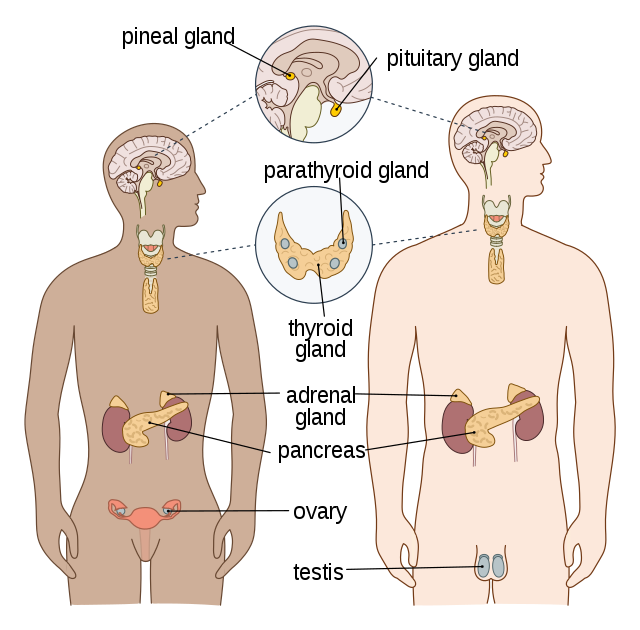
THYMUS GLAND
The thymus gland, situated between the lungs behind the sternum on the ventral side of the aorta, is pivotal in immune system development. Key points include:
- Location:
- The thymus is a lobular structure positioned behind the sternum, between the lungs on the ventral side of the aorta.
- Role in Immune System:
- The thymus plays a crucial role in the development of the immune system.
- Hormones Secretion:
- The thymus gland secretes peptide hormones known as thymosins.
- Function of Thymosins:
- T-Lymphocyte Differentiation: Thymosins are instrumental in the differentiation of T-lymphocytes, contributing to cell-mediated immunity.
- Promotion of Antibody Production: Thymosins also stimulate the production of antibodies, facilitating humoral immunity.
- Age-Related Changes:
- The thymus undergoes degeneration in older individuals, leading to a decline in thymosin production.
- This degeneration results in weakened immune responses in elderly individuals.
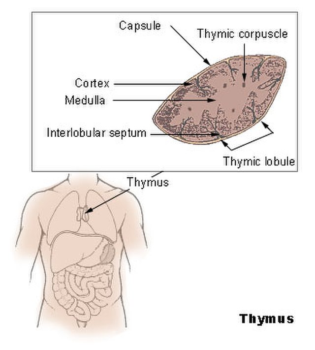
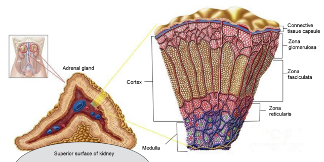
ADRENAL GLAND
The adrenal gland, a pair situated at the anterior part of each kidney, comprises two distinct tissues: the adrenal medulla and the adrenal cortex.
- Location:
- One pair of adrenal glands is located at the anterior part of each kidney.
- Adrenal Medulla:
- The central tissue of the adrenal gland is called the adrenal medulla.
- Adrenal Cortex:
- Surrounding the adrenal medulla is the adrenal cortex.
- Addison’s Disease:
- Underproduction of hormones by the adrenal cortex leads to Addison’s disease.
- Alters carbohydrate metabolism, causing acute weakness and fatigue.
- Adrenal Medulla Hormones (Catecholamines):
- Adrenaline (Epinephrine) and Noradrenaline (Norepinephrine):
- Secreted in response to stress and emergency situations.
- Known as emergency hormones or hormones of Fight or Flight.
- Increase alertness, pupil dilation, piloerection, and sweating.
- Raise heart rate, strengthen heart contractions, and accelerate respiration.
- Stimulate glycogen breakdown, increasing blood glucose.
- Promote lipid and protein breakdown.
- Adrenaline (Epinephrine) and Noradrenaline (Norepinephrine):
- Adrenal Cortex Layers:
- Zona Reticularis (Inner Layer), Zona Fasciculata (Middle Layer), Zona Glomerulosa (Outer Layer):
- Three layers of the adrenal cortex.
- Zona Reticularis (Inner Layer), Zona Fasciculata (Middle Layer), Zona Glomerulosa (Outer Layer):
- Corticoids:
- Hormones secreted by the adrenal cortex, are commonly known as corticoids.
- Glucocorticoids:
- Main Hormone: Cortisol
- Stimulate gluconeogenesis, lipolysis, and proteolysis.
- Inhibit amino acid uptake and utilization.
- Maintain cardiovascular and kidney functions.
- Produce anti-inflammatory reactions and suppress the immune response.
- Main Hormone: Cortisol
- Mineralocorticoids:
- Main Hormone: Aldosterone
- Act on renal tubules, promoting sodium and water reabsorption.
- Excrete potassium and phosphate ions.
- Maintain electrolyte balance, body fluid volume, osmotic pressure, and blood pressure.
- Main Hormone: Aldosterone
- Androgenic Steroids:
- Small amounts are secreted by the adrenal cortex during puberty.
- Influence the growth of axial, pubic, and facial hair.
PANCREAS
The pancreas, a composite gland, functions as both an exocrine and endocrine gland. The endocrine part consists of ‘Islets of Langerhans,’ comprising about 1 to 2 million islets, representing 1 to 2 percent of the pancreas. The two main cell types in the islets are α-cells and β-cells.
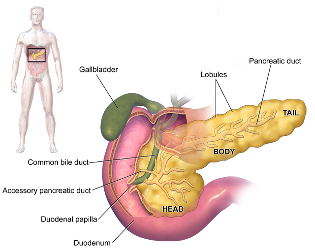
- Islets of Langerhans:
- Located in the endocrine part of the pancreas.
- Contains α-cells and β-cells.
- α-cells:
- Secrete glucagon, a peptide hormone.
- β-cells:
- Secrete insulin, a peptide hormone.
- Glucagon:
- Action:
- Acts on liver cells (hepatocytes).
- Stimulates glycogenolysis, increasing blood sugar (hyperglycemia).
- Stimulates gluconeogenesis, contributing to hyperglycemia.
- Reduces cellular glucose uptake and utilization.
- Role:
- Hyperglycemic hormone.
- Action:
- Insulin:
- Action:
- Acts on hepatocytes and adipocytes.
- Enhances cellular glucose uptake and utilization.
- Promotes rapid movement of glucose from blood to cells, decreasing blood glucose levels (hypoglycemia).
- Stimulates glycogenesis (conversion of glucose to glycogen) in target cells.
- Role:
- Maintains glucose homeostasis.
- Note:
- Works in conjunction with glucagon.
- Action:
- Glucose Homeostasis:
- Jointly maintained by insulin and glucagon.
- Diabetes Mellitus:
- Result of Prolonged Hyperglycemia:
- Loss of glucose through urine.
- Formation of harmful ketone bodies.
- Treatment:
- Successfully treated with insulin therapy.
- Result of Prolonged Hyperglycemia:
TESTIS
The testis, a vital male reproductive organ, serves both as a primary sex organ and an endocrine gland. Positioned in the scrotal sac outside the abdomen, the testis is composed of seminiferous tubules and stromal or interstitial tissue. The Leydig cells or interstitial cells, found in the intertubular spaces, are responsible for producing androgens, primarily testosterone.
- Structure:
- A pair of testis in the scrotal sac.
- Functions:
- Primary Sex Organ:
- Involved in sperm production (spermatogenesis).
- Endocrine Gland:
- Produces androgens, mainly testosterone.
- Primary Sex Organ:
- Androgens (Mainly Testosterone):
- Role in Male Reproductive System:
- Regulate development, maturation, and functions of male accessory sex organs (epididymis, vas deferens, seminal vesicles, prostate gland, urethra).
- Stimulate muscular growth.
- Promote the growth of facial and axillary hair.
- Influence aggressiveness and voice pitch.
- Role in Spermatogenesis:
- Play a major stimulatory role in spermatogenesis.
- Neuroendocrine Effects:
- Act on the central nervous system.
- Influence male sexual behavior (libido).
- Role in Male Reproductive System:
- Metabolic Effects:
- Produce anabolic (synthetic) effects on protein and carbohydrate metabolism.
The testis, with its dual role as a reproductive and endocrine organ, plays a crucial part in male sexual development, secondary sexual characteristics, and overall reproductive functions. The production of testosterone by Leydig cells contributes to various aspects of male physiology and behavior.
OVARY
In the female reproductive system, the ovaries play a pivotal role as the primary sex organ. Positioned in the abdomen, females have a pair of ovaries. The ovary not only produces ova (eggs) during each menstrual cycle but also generates two crucial groups of steroid hormones: estrogen and progesterone.
- Structure:
- A pair of ovaries located in the abdomen.
- Functions:
- Primary Sex Organ:
- Produces ova (eggs) during each menstrual cycle.
- Primary Sex Organ:
- Steroid Hormones:
- Estrogen:
- Synthesized and secreted mainly by growing ovarian follicles.
- Actions include stimulation of:
- Growth and activities of female secondary sex organs.
- Development of ovarian follicles.
- The appearance of female secondary sex characters (e.g., the high pitch of voice).
- Mammary gland development.
- Regulation of female sexual behavior.
- Progesterone:
- Mainly secreted by the corpus luteum (formed after ovulation).
- Supports pregnancy.
- Acts on mammary glands, stimulating:
- Formation of alveoli (milk-storing sac-like structures).
- Milk secretion.
- Estrogen:
- Reproductive Cycle:
- Ovulation: Release of a mature egg from the ovary.
- Corpus Luteum Formation: After ovulation, the ruptured follicle transforms into the corpus luteum, which secretes progesterone.
- Role in Pregnancy:
- Progesterone supports and maintains pregnancy.
- Mammary Gland Development:
- Estrogens and progesterone play roles in the development of mammary glands and milk-related structures.
Ovaries, through the production of estrogen and progesterone, orchestrate various aspects of the female reproductive system, influencing menstrual cycles, pregnancy, and the development of secondary sexual characteristics.
Hormones of Heart, Kidney, and Gastrointestinal Tract
- Heart Hormone – Atrial Natriuretic Factor (ANF):
- Source: Atrial wall of the heart.
- Function: Decreases blood pressure.
- Action: Causes dilation of blood vessels.
- Kidney Hormone – Erythropoietin:
- Source: Juxtaglomerular cells of the kidney.
- Function: Stimulates erythropoiesis (formation of RBC).
- Gastrointestinal Tract Hormones:
- Gastrin:
- Source: Gastric glands.
- Function: Stimulates secretion of hydrochloric acid and pepsinogen.
- Secretin:
- Source: Endocrine cells in the gastrointestinal tract.
- Function: Stimulates secretion of water and bicarbonate ions from the exocrine pancreas.
- Cholecystokinin (CCK):
- Source: Endocrine cells in the gastrointestinal tract.
- Function: Stimulates secretion of pancreatic enzymes and bile juice from the pancreas and gall bladder, respectively.
- Gastric Inhibitory Peptide (GIP):
- Source: Endocrine cells in the gastrointestinal tract.
- Function: Inhibits gastric secretion and motility.
- Gastrin:
- Other Non-Endocrine Tissues:
- Secretion: Growth factors.
- Function: Essential for normal tissue growth, repair, and regeneration.
Mechanism of Hormone Action
- Hormone Receptors:
- Types: Membrane-bound receptors and Intracellular receptors (mainly nuclear receptors).
- Location: Membrane-bound receptors on the cell membrane, Intracellular receptors inside the target cell.
- Formation of Hormone-Receptor Complex:
- Process: Hormones bind to specific receptors, forming hormone-receptor complexes.
- Specificity: Each receptor is specific to a particular hormone.
- Biochemical Changes and Regulation:
- Effect: Formation of hormone-receptor complex induces biochemical changes in the target tissue.
- Regulation: Regulates target tissue metabolism and physiological functions.
- Chemical Nature of Hormones:
- Groups:
- Peptide, Polypeptide, Protein Hormones (e.g., insulin, glucagon, pituitary hormones).
- Steroids (e.g., cortisol, testosterone, estradiol, progesterone).
- Iodothyronines (thyroid hormones).
- Amino-acid derivatives (e.g., epinephrine).
- Groups:
- Interaction with Receptors:
- Membrane-bound Receptors: Interact with receptors on the cell membrane, generating second messengers (e.g., cyclic AMP, IP3, Ca++) to regulate cellular metabolism.
- Intracellular Receptors: Interact with receptors inside the target cell, often regulating gene expression or chromosome function through interaction with the genome.
- Physiological and Developmental Effects:
- Result: Cumulative biochemical actions lead to diverse physiological and developmental effects.