
Types of Movement in Human Cells
- Amoeboid Movement:
- Definition: Movement exhibited by specialized cells like macrophages and leucocytes, resembling Amoeba’s motion.
- Mechanism:
- Pseudopodia formation through streaming of protoplasm.
- Involvement of cytoskeletal elements, particularly microfilaments.
- Ciliary Movement:
- Occurrence: In internal tubular organs with ciliated epithelium.
- Examples:
- Coordinated cilia movements in the trachea aid in removing inhaled dust particles.
- Facilitates the passage of ova through the female reproductive tract.
- Muscular Movement:
- Role: Essential for various body movements, including limb, jaw, and tongue actions.
- Contractile Property: Muscles contract to generate force, enabling locomotion and other bodily movements.
- Coordination: Requires synchronized activity of muscular, skeletal, and neural systems.
Muscle: Structure and Classification
- Overview:
- Muscle is a specialized tissue of mesodermal origin.
- Constitutes 40-50% of the body weight in humans.
- Possesses unique properties: excitability, contractility, extensibility, and elasticity.
- Muscle Classification:
- Based on Location:
- Skeletal Muscles:
- Associated with skeletal components.
- Striped appearance (striated muscles).
- Voluntary control by the nervous system.
- Involved in locomotion and posture changes.
- Visceral Muscles:
- Found in the inner walls of visceral organs.
- Smooth appearance (smooth muscles or nonstriated).
- Involuntary control.
- Functions in the transport of food and gametes.
- Cardiac Muscles:
- Found in the heart.
- Striated appearance.
- Involuntary nature; The nervous system indirectly regulates.
- Skeletal Muscles:
- Based on Location:
- Structure of Skeletal Muscle:
- Organized into muscle bundles or fascicles held by fascia.
- The muscle bundle contains muscle fibers.
- Muscle fiber: sarcolemma, sarcoplasm, and multiple nuclei.
- The sarcoplasmic reticulum stores calcium ions.
- Myofibrils or myofilaments (actin and myosin) give the striated appearance.
- Sarcomere: Functional unit of contraction between two ‘Z’ lines.
- Myofibril Structure:
- Actin and Myosin:
- Light bands (I-band) contain actin.
- Dark bands (A-band) contain myosin.
- Actin and myosin are arranged parallel to each other.
- ‘Z’ line bisects the ‘I’ band; thin filaments attached to ‘Z’ line.
- ‘M’ line in the ‘A’ band holds thick filaments together.
- Sarcomere: Functional unit between two ‘Z’ lines.
- Resting State:
- Thin filaments partially overlap the free ends of thick filaments.
- A central part of the thick filament not overlapped is the ‘H’ zone.
- Actin and Myosin:
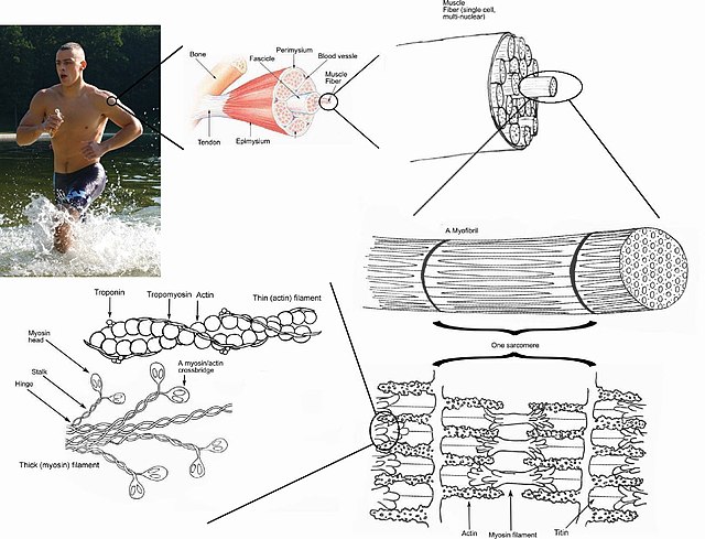
Structure of Contractile Proteins
- Actin Filament (Thin Filament):
- Composed of two helically wound ‘F’ (Filamentous) actins.
- Each ‘F’ actin is a polymer of monomeric ‘G’ (Globular) actins.
- Tropomyosin, another protein, runs close to ‘F’ actins.
- Troponin, a complex protein, is distributed at regular intervals on tropomyosin.
- In the resting state, the troponin subunit masks active binding sites for myosin on actin filaments.
- Myosin Filament (Thick Filament):
- A polymerized protein consisting of monomeric proteins called Meromyosins.
- Each Meromyosin has a globular head with a short arm (Heavy Meromyosin – HMM) and a tail (Light Meromyosin – LMM).
- The globular head, known as the cross arm, projects outwards at regular intervals.
- The globular head is an active ATPase enzyme with binding sites for ATP and active sites for actin.
Mechanism of Muscle Contraction
- Sliding Filament Theory:
- Muscle contraction involves the sliding of thin filaments over thick filaments.
- Initiated by a neural signal from the central nervous system (CNS) via a motor neuron.
- Motor Unit:
- Motor neurons and connected muscle fibers form a motor unit.
- The neuromuscular junction or motor-end plate is the junction between a motor neuron and the muscle fiber’s sarcolemma.
- Neuromuscular Junction Activation:
- Neural signal releases acetylcholine, generating an action potential in the sarcolemma.
- Action potential spreads through the muscle fiber, releasing calcium ions into the sarcoplasm.
- Calcium Ion Binding:
- Calcium binds with troponin on actin filaments, exposing active sites for myosin.
- Myosin head binds to exposed active sites, forming a cross-bridge.
- Cross-Bridge Formation:
- Myosin pulls attached actin filaments towards the center of ‘A’ band, shortening the sarcomere (contraction).
- ‘Z’ line attached to actins is also pulled inwards during contraction.
- Cross-Bridge Breakage:
- Myosin releases ADP and P1, returning to its relaxed state.
- ATP binds, breaking the cross-bridge.
- ATP hydrolysis by myosin head repeats the cycle, causing further sliding.
- Relaxation:
- Ca++ ions are pumped back to sarcoplasmic cisternae, masking actin filaments.
- ‘Z’ lines return to their original position, causing relaxation.

SKELETAL SYSTEM

- Framework of Bones and Cartilages:
- Essential for body movement.
- Bone and cartilage are specialized connective tissues.
- The bone matrix contains hard calcium salts, and cartilage has pliable chondroitin salts.
- Composition:
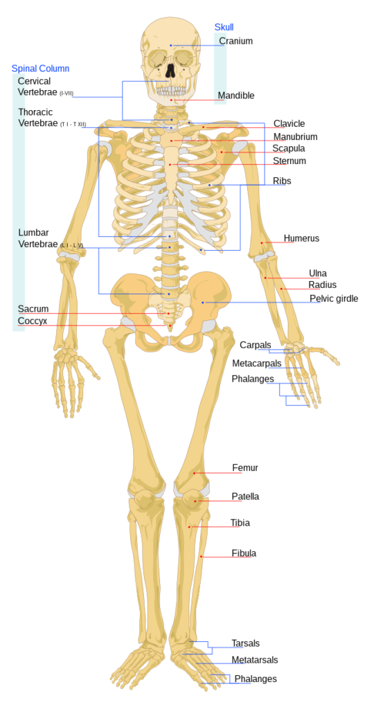
- 206 bones and a few cartilages in humans.
- Divided into axial and appendicular skeleton.
- Axial Skeleton (80 Bones):
- Skull: Cranial (8 bones) and facial (14 bones) elements.
- Hyoid: U-shaped bone at the base of the buccal cavity.
- Ear Ossicles: Malleus, Incus, Stapes in each middle ear.
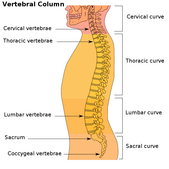
- Vertebral Column (26): Cervical (7), thoracic (12), lumbar (5), sacral (1 fused), coccygeal (1 fused).
- Sternum: Flat bone on the ventral midline of the thorax.
- Ribs (12 pairs): True ribs (1-7), false ribs (8-10), floating ribs (11-12).
- Appendicular Skeleton:
- Limb Bones (30 in each limb): Humerus, radius, ulna, carpals (8), metacarpals (5), phalanges (14).
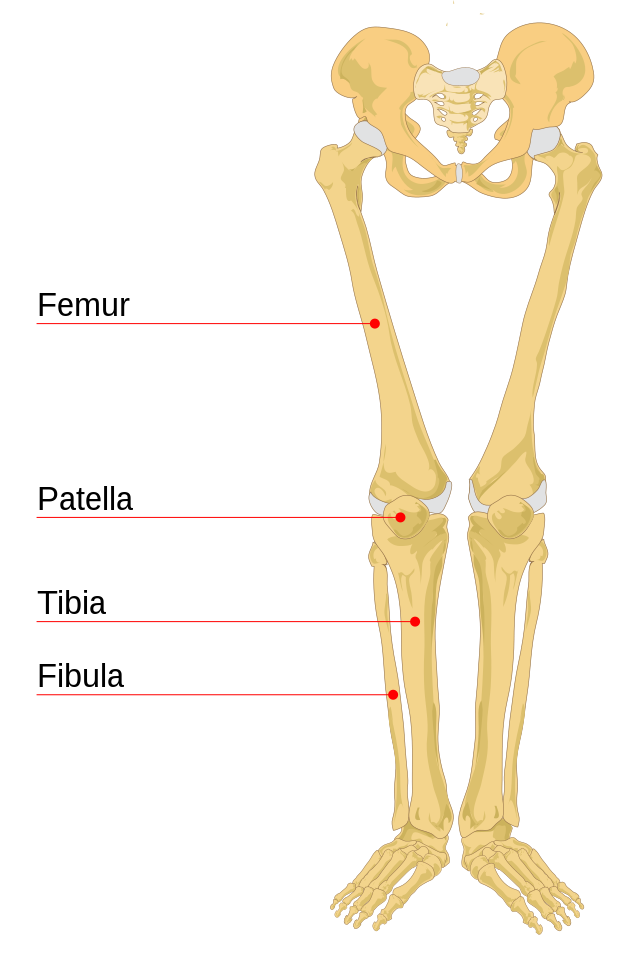
- Thigh Bone: Femur.
- Leg Bones: Tibia, fibula, tarsals (7), metatarsals (5), phalanges (14).
- Knee Cap: Patella.
- Girdles:
- Pectoral Girdle: Clavicle and scapula on each side. Articulates with the humerus and forms shoulder joint.
- Pelvic Girdle: Coxal bones (fusion of ilium, ischium, and pubis). Articulates with the thigh bone.
- Articulation:
- Pectoral and pelvic girdles allow articulation of upper and lower limbs with the axial skeleton.
JOINTS
- Functionality:
- Essential for all types of body movements, including locomotion.
- Joints serve as points of contact between bones or between bones and cartilage.
- Muscular force is applied at joints, acting as fulcrums for movement.
- Classification:
- Fibrous Joints:
- No movement allowed.
- Example: Flat skull bones fuse end-to-end with dense fibrous connective tissues (sutures) forming the cranium.
- Cartilaginous Joints:
- Bones joined by cartilages.
- Example: Joint between adjacent vertebrae in the vertebral column, allowing limited movements.
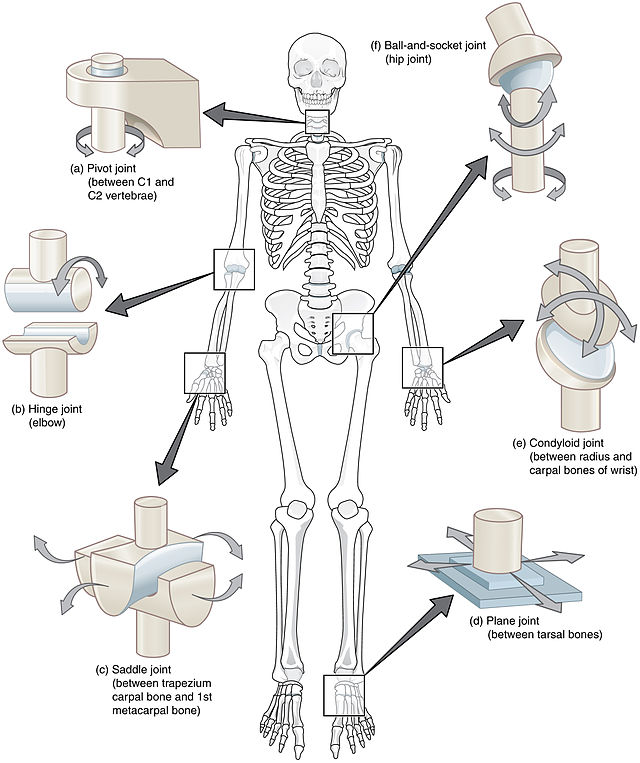
- Synovial Joints:
- Characterized by a fluid-filled synovial cavity between articulating surfaces.
- Allows considerable movement.
- Examples: Ball and socket joint (humerus and pectoral girdle), hinge joint (knee), pivot joint (atlas and axis), gliding joint (between carpals), saddle joint (between carpal and metacarpal of the thumb).
- Fibrous Joints:
- Joint Movements:
- Ball and Socket Joint:
- Example: Between humerus and pectoral girdle.
- Hinge Joint:
- Example: Knee joint.
- Pivot Joint:
- Example: Between atlas and axis.
- Gliding Joint:
- Example: Between carpals.
- Saddle Joint:
- Example: Between carpal and metacarpal of the thumb.
- Ball and Socket Joint:
DISORDERS OF THE MUSCULAR AND SKELETAL SYSTEM
- Myasthenia Gravis:
- Definition: Autoimmune disorder affecting the neuromuscular junction.
- Effects: Leads to fatigue, weakening, and paralysis of skeletal muscles.
- Muscular Dystrophy:
- Definition: Progressive degeneration of skeletal muscles, often of genetic origin.
- Tetany:
- Description: Rapid spasms (wild contractions) in muscles.
- Cause: Low calcium levels in body fluids.
- Arthritis:
- Description: Inflammation of joints.
- Characteristics: Pain, swelling, and reduced joint mobility.
- Osteoporosis:
- Definition: Age-related disorder characterized by decreased bone mass.
- Consequences: Increased susceptibility to fractures.
- Common Cause: Decreased levels of estrogen.
- Gout:
- Description: Inflammation of joints.
- Cause: Accumulation of uric acid crystals.
- Symptoms: Joint pain and swelling.