COORDINATION IN THE HUMAN BODY
Coordination is essential for maintaining homeostasis, ensuring that the functions of various organs and organ systems work harmoniously. This process involves the interaction and complementation of different organs to achieve a balanced state. For instance, during physical exercises:
- Increased Energy Demand:
- Effect: Muscular activity rises, increasing the demand for energy.
- Response: Oxygen supply is enhanced.
- Oxygen Supply and Respiratory Rate:
- Effect: Increased need for oxygen.
- Response: Respiration rate escalates, ensuring higher oxygen intake.
- Cardiovascular System:
- Effect: Enhanced oxygen requirement and removal of metabolic by-products.
- Response: Increased heart rate and blood flow through blood vessels.
- Post-Exercise Recovery:
- Effect: Activities of nerves, lungs, heart, and kidneys gradually return to normal.
- Process: Coordinated return to baseline conditions.
Coordination Systems:
- Neural System:
- Description: Organized network of point-to-point connections.
- Advantage: Quick coordination.
- Endocrine System:
- Description: Chemical integration through hormones.
- Advantage: Provides sustained and widespread coordination.
Integration of Neural and Endocrine Systems:
- Objective: Ensure synchronized functioning of organs.
- Role: Coordination and integration of activities for optimal physiological performance.
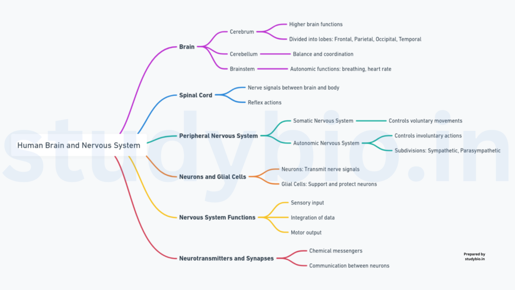
NEURAL SYSTEM IN HUMANS
The neural system, fundamental for coordination, is composed of specialized cells known as neurons. Neurons are capable of detecting, receiving, and transmitting diverse stimuli. The neural organization varies across different organisms.
Neural System in Lower Invertebrates:
- Example: Hydra
- Description: Simple network of neurons.
Neural System in Insects:
- Features: Brain, ganglia, and neural tissues present.
Human Neural System:
- Central Neural System (CNS):
- Components: Brain and spinal cord.
- Function: Site for information processing and control.
- Peripheral Neural System (PNS):
- Components: Nerves associated with CNS.
- Types of Nerve Fibres:
- (a) Afferent Fibres: Transmit impulses to CNS from tissues/organs.
- (b) Efferent Fibres: Transmit regulatory impulses from CNS to peripheral tissues/organs.
Subdivisions of PNS:
- Somatic Neural System:
- Function: Relays impulses from CNS to skeletal muscles.
- Autonomic Neural System:
- Function: Transmits impulses from CNS to involuntary organs and smooth muscles.
- Subdivisions:
- (a) Sympathetic Neural System
- (b) Parasympathetic Neural System
Visceral Nervous System:
- Definition: Part of PNS encompassing nerves, fibers, ganglia, and plexuses.
- Function: Facilitates impulse travel between the central nervous system and viscera.
NEURON: STRUCTURAL AND FUNCTIONAL UNIT
A neuron, the fundamental building block of the neural system, is a microscopic structure comprising three essential parts:
- Cell Body:
- Components: Cytoplasm, cell organelles, Nissl’s granules.
- Function: Core structure housing essential cellular components.
- Dendrites:
- Characteristics: Short fibers branching from the cell body.
- Contains: Nissl’s granules.
- Function: Transmit impulses toward the cell body.
- Axon:
- Structure: Long fiber with branched distal end.
- Termination: Bulb-like synaptic knobs.
- Contains: Synaptic vesicles with neurotransmitters.
- Function: Transmit nerve impulses away from the cell body to synapses or neuro-muscular junctions.
Types of Neurons Based on Axons and Dendrites:
- Multipolar Neurons:
- Description: One axon and two or more dendrites.
- Location: Found in the cerebral cortex.
- Bipolar Neurons:
- Description: One axon and one dendrite.
- Location: Found in the retina of the eye.
- Unipolar Neurons:
- Description: Cell body with one axon only.
- Location: Commonly found in the embryonic stage.
Types of Axons:
- Myelinated Axons:
- Surrounding: Enveloped with Schwann cells forming a myelin sheath.
- Characteristics: Nodes of Ranvier present between adjacent myelin sheaths.
- Location: Found in spinal and cranial nerves.
- Unmyelinated Axons:
- Surrounding: Enclosed by Schwann cells without forming a myelin sheath.
- Location: Commonly found in both autonomous and somatic neural systems.
GENERATION AND CONDUCTION OF NERVE IMPULSE
Neurons exhibit excitability due to their polarized membrane state. The polarization arises from selective ion channels on the neural membrane, with potassium ions (K+) being more permeable and sodium ions (Na+) nearly impermeable during the resting state. This is maintained by the sodium-potassium pump actively transporting ions.
Resting Potential:
- Condition: Neuron at rest.
- Characteristics: Axonal membrane permeable to K+, impermeable to Na+.
- Result: Positive charge on the outer surface, negative charge on the inner surface, creating a polarized state.
Stimulus-Induced Changes:
- Stimulus: Applied at a specific site (e.g., point A).
- Response: The membrane at site A becomes permeable to Na+, causing a rapid influx.
- Outcome: Reversal of polarity, outer surface negatively charged, inner surface positively charged (depolarization).
Action Potential (Nerve Impulse):
- Definition: Rapid reversal of membrane polarity at the stimulated site.
- Propagation: Sequential depolarization at sites along the axon.
- Conduction: Current flow from site to site, completing the circuit.
Conduction Mechanism:
- Depolarization at Site A:
- The membrane becomes permeable to Na+.
- The rapid influx of Na+.
- Reversal of polarity at site A.
- Generation of action potential.
- Propagation to Site B:
- Site B membrane has the opposite polarity.
- Current flows on inner and outer surfaces.
- Polarity reversal at site B.
- Generation of action potential at site B.
- Sequential Impulse Propagation:
- Repetition along the axon.
- Current flow maintains impulse conduction.
Termination of Action Potential:
- Process: Short-lived rise in Na+ permeability.
- Outcome: Followed by a rise in K+ permeability.
- Result: K+ diffusion outside the membrane.
- Effect: Restores resting potential, making the fiber responsive to further stimulation.
TRANSMISSION OF IMPULSES
A nerve impulse travels between neurons through specialized junctions called synapses. Synapses can be classified into two types: electrical synapses and chemical synapses.
- Electrical Synapses:
- Description: Close proximity of pre-and post-synaptic neuron membranes.
- Transmission: Direct flow of electrical current from one neuron to another.
- Speed: Faster compared to chemical synapses.
- Occurrence: Rare in the human system.
- Chemical Synapses:
- Structure: Membranes of pre-and post-synaptic neurons separated by a fluid-filled synaptic cleft.
- Transmission: Involves neurotransmitters.
- Axon Terminals: Contain vesicles filled with neurotransmitters.
The process at Chemical Synapses:
- Impulse Arrival:
- Stimulus: Arrival of an impulse (action potential) at the axon terminal.
- Response: Stimulation of synaptic vesicle movement towards the membrane.
- Neurotransmitter Release:
- Action: Synaptic vesicles fuse with the plasma membrane.
- Outcome: Release of neurotransmitters into the synaptic cleft.
- Post-Synaptic Binding:
- Interaction: Neurotransmitters bind to specific receptors on the post-synaptic membrane.
- Result: Opening of ion channels on the post-synaptic membrane.
- Ion Entry and New Potential:
- Effect: Entry of ions through open channels.
- Outcome: Generation of a new potential in the post-synaptic neuron.
- Nature: Potential can be excitatory or inhibitory.
CENTRAL NEURAL SYSTEM
The central neural system (CNS) is the hub of information processing and control in the human body, primarily composed of the brain and the spinal cord. The brain, as the central command center, orchestrates various functions and activities, both voluntary and involuntary.
- Brain Functions:
- Control: Regulates voluntary movements, body balance, and vital involuntary organ functions (e.g., lungs, heart, kidneys).
- Regulation: Manages thermoregulation, hunger, thirst, and circadian rhythms.
- Processing: Handles sensory information for vision, hearing, and speech.
- Cognition: Processes memory, intelligence, emotions, and thoughts.
- Behavior: Influences human behavior.
- Brain Protection:
- Cranial Meninges: Layers covering the brain inside the skull.
- Dura Mater: Outer layer.
- Cranial Meninges: Layers covering the brain inside the skull.
- Brain Divisions:
- (i) Forebrain:
- Functions: Complex processing, sensory integration, and higher-order cognitive functions.
- Components: Cerebrum (largest part), thalamus, and hypothalamus.
- (ii) Midbrain:
- Location: Between forebrain and hindbrain.
- Functions: Relay station for auditory and visual information.
- (iii) Hindbrain:
- Functions: Vital processes, coordination of motor activities.
- Components: Cerebellum, pons, and medulla oblongata.
- (i) Forebrain:
FOREBRAIN
The forebrain, a crucial part of the central neural system, comprises the cerebrum, thalamus, and hypothalamus. Each component plays a distinct role in various physiological and cognitive functions.
- Cerebrum:
- Major Component: Forms the largest part of the human brain.
- Structural Division: Divided into left and right cerebral hemispheres by a deep cleft.
- Connectivity: Hemispheres connected by the corpus callosum, a tract of nerve fibers.
- Cerebral Cortex: Outer layer with prominent folds (gyri and sulci).
- Grey Matter: Neuron cell bodies are concentrated, giving a greyish appearance.
- White Matter: Inner part of cerebral hemispheres, composed of myelinated nerve fibers, giving an opaque white appearance.
- Thalamus:
- Position: Central coordinating center, wrapped around by the cerebrum.
- Function: Major role in sensory and motor signaling.
- Hypothalamus:
- Location: Situated at the base of the thalamus.
- Functions:
- Regulates body temperature.
- Complex Structure: Includes inner parts of cerebral hemispheres and associated structures (amygdala, hippocampus) forming the limbic lobe or limbic system.
- Involvement: Regulates sexual behavior, emotional reactions (excitement, pleasure, rage, fear), and motivation.
MIDBRAIN
The midbrain, positioned between the thalamus/hypothalamus of the forebrain and the pons of the hindbrain, plays a vital role in the central neural system. Its distinct features include the presence of the cerebral aqueduct and four round swellings known as corpora quadrigemina.
- Location:
- Position: Between the thalamus/hypothalamus (forebrain) and pons (hindbrain).
- Passageway: Contains the cerebral aqueduct, a canal traversing through the midbrain.
- Corpora Quadrigemina:
- Structure: Dorsal portion characterized by four round swellings.
- Functions: Involved in visual and auditory reflexes.
- Superior Colliculi: Upper pair, associated with visual reflexes.
- Inferior Colliculi: Lower pair, involved in auditory reflexes.
HINDBRAIN
The hindbrain, a crucial division of the brain, encompasses the pons, cerebellum, and medulla oblongata. Each component serves distinct functions in maintaining vital bodily processes.
- Pons:
- Composition: Consists of fiber tracts facilitating interconnection across various brain regions.
- Function: Facilitates communication between different parts of the brain.
- Cerebellum:
- Surface: Highly convoluted surface, maximizing space for an abundance of neurons.
- Function: Plays a key role in motor control and coordination.
- Medulla (Medulla Oblongata):
- Connection: Links to the spinal cord.
- Centers: Contains centers regulating respiration, cardiovascular reflexes, and gastric secretions.
Brain Stem:
- Constituents: Comprises the midbrain, pons, and medulla oblongata.
- Function: Establishes connections between the brain and spinal cord.